Spore trap analysis [ST]
Published: July 8th, 2009
Revised: March 24th, 2023
Spore trap sampling involves collecting airborne particles on a filter membrane or adhesive-coated slide by drawing air through or over the collection medium, respectively. The collection medium is then analyzed by transmitted light microscopy, usually at 600–1000× magnification. A number of different collection devices may be used for spore trap sampling of which the most common are 1) slit impactors such as the Allergenco-D or Air-O-Cell cassette, and 2) mixed cellulose ester membrane (MCEM) filters.
The normally high minute-to-minute variability in levels of airborne spores greatly limits the utility of short-term quantitative air sampling data. This large intrinsic source of variation also minimizes the importance of counting method as a significant source of error. The most critical source of error in spore trap analysis qualitative, involving the identification of spores. Spore trap analysis is one of the most technically challenging tasks in environmental mycology, requiring considerable skill and experience on the part of the analyst, to identify spores accurately, and to differentiate them from other airborne particles. A good spore trap analyst requires years of experience not just analyzing spore trap samples but examining whole specimens from field collections in order to learn the full variation of fungal structures as well as particles that might be confused with them. This level of experience cannot be trained in a short time, and it cannot easily be acquired by individuals lacking advanced training in mycology or botany. All of our analysts have extensive, supervised experience in fungal spore identification. They participate regularly in proficiency testing programs through the American Industrial Hygiene Association, Proficiency Analytical Testing Programs (AIHA-PAT, LLC), and they have attained independent certification of their skills through proctored examinations by the Pan–American Aerobiology Certification Board. For more information on the requirements visit the PAN-AMERICAN AEROBIOLOGY CERTIFICATION BOARD >
Spore trap sampling has an advantage over culture-dependent methods by permitting the enumeration of fungal particles regardless of culturability or viability. Because of this, spore trap sampling is sometimes incorrectly referred to as a “non-viable” sampling method; however, it more appropriately should be called a “total” sampling method, since both culturable as well as non-culturable propagules are counted. The greatest limitation of spore trap sampling is its inability to allow low-level identifications of spores due to the lack of diagnostic features on many spore types. By contrast, culture-dependent methods have the advantage of access to a variety of cell types for a given fungus, as well as a living culture that could be submitted to physiological tests, allowing other distinguishing characteristics to be evaluated. As such, spores counted in spore traps can only be identified presumptively based on their size, shape and color.
Method 1. Slit impactor sampling
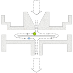 Disposible slit impactor samplers consist of a plastic housing with a tapered slit-like window on one side and an outlet tube on the other side. In the interior of the cassette, a small glass or plastic wafer is positioned slightly below the open window of the slit. The wafer is typically coated with an optically clear, sticky gel or grease. As air is drawn through the slit, particles are drawn towards the wafer. Particles of relatively small momentum will deflect with the air stream and be exhausted on the outlet side of the cassette. Particles of relatively larger momentum may be incompletely deflected at the wafer surface, and impact on its sticky surface. The size of particles captured with 50% efficiency on the wafer is referred to as the d50 cutpoint of the sampler. Particles larger than the d50 cutpoint are collected with very high efficiency, whereas particles smaller than the d50 cutpoint are poorly collected by the sampler. Most modern single-use slit samplers are individually serial-numbered and stamped with an expiry date, indicating the usable life of their adhesive and assembly.
Field sample collection
- Calibrate a high flow air sampling pump to collect 15-20 L/min using an equivalent calibration cassette placed in-line. The selection of actual flow rate is important, since collection efficiency is lower and the particle deposit tends to become more diffuse at lower collection flow rates. While higher flow rates are generally better from the standpoint of impaction efficiency, increased air velocities tend to increase particle bounce, which may result in loss. Most manufacturers of single-use slit impactors recommend sample collection at a flow rate of 15 L/min.
- Verify that the collection medium has a current expiration date.
- Connect the vacuum hose to the outlet of the cassette. Support the cassette or hose so that the sample slit is oriented in a downward direction. Remove the slit cover.
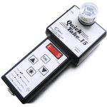 Most manufacturers recommend sampling volumes of 150 L for generally clean indoor environments and 75 L for environments suspected to be overly dusty, or for the collection of outdoor comparison samples. Sampling volumes lower than this (e.g. 10-25 L) may be helpful in areas that are highly dusty, such as remediation enclosures or dust-generating industrial processes.
- At least one field blank should be collected for every 10 field samples taken. Field blanks should be handled in exactly the same manner as test samples except that air must not be drawn through them.
- Following collection, samples and blanks should be re-sealed, labeled and arranged in a clean box or bag away from moisture and light until analysis.
- Calibrate a high-volume pump with an calibration filter cassette in-line to achieve a flow rate between 2-15 L/min air.
- Connect the vacuum hose to the outlet nipple of the cassette and remove the end-cap from the front of the cassette. Samples should be collected with the cassette oriented downward in order to reduce over-sampling from gravity-related particle deposition. Ideally, between 1.0-1.2 m³ air should be collected, although lower total air volumes may be indicated in dusty atmospheres.
- One field blank handled exactly as test samples (without any air draw) should be analyzed for every 10 field samples.
- After collection, all cassettes should be sealed, labeled and placed in a clean box or bag away from light and moisture until analysis.
- Collect a 1–2 g sample of bulk material using a clean knife, chisel, or other implement suitable for the material in question. The collection tools should be sterilized by saturating the surfaces with 70% isopropyl alcohol (the surfaces should remain damp for at least 2 min) prior to use.
- If the sample is dry, place it in a sturdy closable plastic bag such as a zippered freezer bag. Damp samples should be placed in a clean new paper bag or envelope in order to prevent fungal growth in transit post collection. Damp samples should be received by the laboratory no later than 24 hr post collection. Dry samples can be retained for a longer period of time prior to analysis without producing spurious results.
- If using optically clear adhesive tape (e.g., 3M Scotch “Red Tartan” Brand tape), strip away and discard the leading 5–10 cm of tape, then remove a 2–5 cm segment for sampling. Tape used for sampling should be kept in a clean reclosable plastic bag and not be used for any other purpose. To control against cross-contamination, one field blank (unsampled tape lift handled similarly to actual field samples) should be taken and analyzed for each ten field samples.
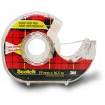 Press the adhesive tape or BioTape against the surface to be tested. The collection of superficial debris from rough or irregular surfaces can be improved by gently rubbing the pad of your index finger against the backing of the tape.
- Replace the BioTape strip in the transport shell. If using adhesive tape, adhere the tape strip to the interior surface of a sturdy closable plastic bag such as a zippered freezer bag (do not fold the tape onto itself).
Method 2. Mixed cellulose ester membrane (MCEM) sampling
MCEM spore traps typically require a longer sampling period than slit-type collectors (e.g. 1–8 hr vs. 0.5–30 min, respectively). The longer-term collection however yields a more integrated, average sample, capturing transient fluxes over time. The spore catch in an MCEM sample tends also to be evenly distributed over the membrane surface, making the analysis of these samples less sensitive to sample overloading. As well, the fact that the entire spore catch lies in a single focal plane makes this sampling method appealing from a laboratory analytical standpoint.
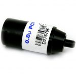 Because fitration-based methods rely much less than slit impactors on particle momentum to capture and retain particles, filter samplers may be used over a wide range of flow rates without suffering appreciable changes in the collection yield. Typically, a 25 mm MCEM filter with 0.8 µm pore size is used for collection. Suitable filters include asbestos PCM cassettes, that are available pre-loaded in 3-piece electrically conductive cassette outfitted with a 50 mm extension cowl that reduces the dispersion of collected particles due to static electric effects. The laboratory analysis of MCEM filter samples involves the use of a “hot block” vaporizer to generate acetone vapour for clearing the filter, followed by application of a microscopic mounting fluid to the cleared surface of the membrane and examination under the compound microscope.
Field sample collection
Laboratory code: ST
Service options

Results reporting
Results of spore trap samples will be reported quantitatively as Elements/m³ for each organism category observed. The background level of non-fungal particles is reported semiquantitatively, as 1+, 2+, 3+, etc., where the numeral preceding the plus sign indicates the level of visible background particles per optical field in accordance with ASTM D7391-09. Background levels above 3+ indicate that the quantitation of fungal particles in the sample may be unreliable due to oversampling.
Bulk sample or tape lift/ BioTape sample for microscopic analysis [BT]
Published: July 8th, 2009
Revised: March 3rd, 2023
Direct microscopy is considered the “gold standard” in environmental mycology for determining the existence of fungal growth on surfaces or materials. The direct observation of fungal spores, spore-bearing structures and mycelium provides an unequivocal marker of past or current growth. Direct microscopic examination of tape or bulk samples should first examine the specimen in reflected light at a magnification of 10–40 × for gross evidence of colonization. After that, preparations from one or more areas of the specimen should be prepared for evaluation by transmitted light microscopy at higher magnification (e.g. 400–1000 ×). There are two basic methods for examining bulk specimens by transmitted light microscopy: 1) adhesive tape lift and 2) bulk scraping/ shaving.
Bulk samples
Bulk samples include 1) solid materials like wood, 2) fibrous materials such as carpet and dust, and 3) powdery materials like drywall. The direct evaluation of bulk samples is informative because the observation of fungal filaments is a conclusive marker of fungal growth and represents an unacceptable condition on indoor materials. The collection of bulk samples is also usually simple in the field, and the examination of these samples is typically straightforward in the laboratory.
Field collection method
Adhesive tape lifts and BioTape samples
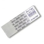 For nearly a century, shellac peels have been used to collect and study foliar diseases of plants. More recently, these methods have been modified to assist in the examination of bulk materials and fixed surfaces from indoor environments for the presence of fungal growth. Adhesive tape methods are more practical than liquid shellacs because they do not require lengthy curing times, they can be used easily on highly porous materials, and they tend not to cause appreciable surface disfigurement. Adhesive tape samples are highly useful in the laboratory because they tend to preserve the arrangement of spores and spore-bearing structures, greatly assisting the process of identification. A number of genera of important indoor fungal contaminants may be recognized directly from spores alone (e.g., Chaetomium, Stachybotrys). Many more genera may be identified if spores as well as spore-bearing structures remain intact.
Tape sampling is useful for 1) field collection of surfaces where fungal colonization is suspected; 2) laboratory sampling of bulk specimens; 3) laboratory preparations from cultures where the preservation of delicate microscopic structures is required for identification. These media have a long shelf life and are very easy to handle in the field.
Field collection method
Laboratory code: BT
Service options

Results reporting
Direct microscopic examination of bulk and tape lift samples allows for the identification and semiquantification of fungal colonists on a surface. Results are normally reported on a genus by genus basis, with the numbers of spore and vegetative filaments described semiquantitatively (e.g., 1+, 2+, 3+, etc.). Fungal spores arising from outdoor materials are common in indoor settled dusts. Therefore, the simple observation of fungal spores on an indoor sample is insufficient to conclude that the surface is a fungal growth site. Usually, this determination requires the microscopic observation of both fungal filaments (vegetative hyphae/ mycelia/ spore-bearing structures) and spores.
Mycobacterium immunogenum in metalworking fluids [MIPCR]
Published: July 8th, 2009
Revised: March 3rd, 2023
Exposure to semisynthetic and synthetic metalworking fluids (MWF) has been associated with work-related asthma, hypersensitivity pneumonitis and dermatitis. Exposure to microbial contaminants in MWFs appears to be a key factor in the etiology of these diseases in machinists. Recently released occupational exposure limits to MWFs are based on aerosol concentrations determine by gravimetric measurements that have failed to consider the relevance of bioaerosol exposure, including viable airborne microbes (bacteria and fungi) and microbe-derived chemicals, such as endotoxin (Gram-negative bacteria) and ergosterol (moulds and yeasts). One bacterium in particular, Mycobacterium immunogenum, was described recently from synthetic and semi-synthetic MWFs. Recent research suggests that this bacterium plays a major role in respiratory disease of workers exposed to microbially contaminated MWFs.
This test detects members of the M. immunogenum complex using PCR. This method cannot distinguish between living and dead cellular material. A negative result does not necessarily refute the presence of potentially infectious material in the area tested, since only a small amount of material is subjected to the test. The testing of multiple replicate samples is helpful to establish confidence on the generalizability of results. A positive result, however, may provide information that is useful in public health interventions.
Bulk samples of MWF should be collected dry in a clean, new, food-grade, bottle. A minimum volume of 100 mL should be collected. Air samples may be collected on a 25 or 37 mm 0.4 um polycarbonate membrane filter. A minimum of 1 cubic metre of air should be collected. Both sample formats should be placed individually in a clean, new, food-grade, zippered freezer bag, and delivered to the laboratory within 24 hr of collection.
Laboratory code: MIPCR
Service options

Endotoxin [ENDO]
Published: July 8th, 2009
Revised: March 3rd, 2023
Endotoxin is a lipopolysaccharide found in the cell membranes of Gram-negative bacteria. Endotoxin is a strongly proinflammatory material because of its interactions with receptors that stimulate the innate immune system. Exposure to endotoxin will initiate a strong inflammatory response in anyone and does not require allergic sensitization or individual sensitivity. Inhalation exposure to endotoxin is thought to be a contributor to a number of inflammatory airways diseases such as Farmer’s Lung Disease and other hypersensitivity pneumonitides.
Endotoxin exposures typically arise where fluids contaminated with Gram negative bacteria become nebulized, such as spray-mist type humidification systems, hot tubs. In industry, sewage treatment processes and synthetic and semisynthetic machining coolants are a common source of endotoxins.
This test measures endotoxin content of air (air samples for endotoxin must be collected using special endotoxin-free polycarbonate filter membranes), bulk or TefTex wipe samples using the Limulus Amoebocyte Lysate (LAL) assay. This sensitive bioassay measures endotoxin by a reaction that uses highly sensitive endotoxin recombinant receptors cloned from blood cells of the horseshoe crab (genus Limulus). This is the gold standard method of measuring endotoxin, and it is used in a wide range of applications including the testing of medical devices and pharmaceutical preparations.
We reported levels in endotoxin units (EU) per standard unit of material tested (e.g. EU/mg, EU/m³, etc.). This measure can be converted to gravimetric units (e.g., ng) if required. In the case of air samples, endotoxin levels in a test area are usually compared to background levels in a non-complaint area. Airborne endotoxin greater than 30 times above background has been suggested as an action-level (ACGIH 1999).
Laboratory code: ENDO
Service options

Bacterial DNA barcode sequence identification [BDNA]
Published: July 8th, 2009
Revised: February 23rd, 2023
This test provides a low-level identification of a single bacterial isolate by sequencing of portions of the 16S ribosomal small subunit gene region. DNA-based sequence identification is the gold-standard for microbial identification, since it is capable of identifying rare or atypical isolates of bacteria irrespective of their responses to diagnostic culture media or biochemical tests.
DNA is isolated from cells using standard procedures. A diagnostic portion of the ribosomal small subunit gene is amplified by PCR and sequenced in both directions. A consensus sequence of both forward and reverse strands is constructed, and this sequence is used to query the Ribosomal Database Project (RDP), an online data analysis and alignment tool consisting of annotated Bacterial and Archaeal small-subunit 16S rRNA sequences. This approach to bacteria identification is unmatched in its ability to accurately identify troublesome isolates, or to ascertain the taxonomic position of novel strains.
Laboratory code: BDNA
Service options


