Bulk/ swab/ wipe, semiquantitative fungal culture [FC-S]
Published: July 8th, 2009
Revised: March 21st, 2023
Bulk samples
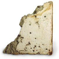 In the laboratory, a semiquantitative fungal culture of a bulk sample is generally carried out by aseptically taking tiny shavings from the specimen and sprinkling them over the surface of a Petri dish filled with growth medium. Alternatively, a swab may be used in the laboratory to recover any loose debris from the surface of a bulk sample, and then rolled on the surface of a growth medium. A third method for semiquantitative testing of bulk samples involves washing all or a portion of the surface of the material with a sterile buffer solution, and transferring an aliquot of the resulting suspension to a Petri dish. Which particular procedure is indicated is a subjective decision and it depends to a great extent on the size, composition and overall appearance of the sample. The advantage to starting with a bulk sample is that it permits the laboratory to determine which approach is most suitable, possibly including several parallel approaches to achieve the most accurate result.
Swab samples
 Despite their ease of handling, swab samples are generally poorly suited as field methods for sample collection. Swabs with a woven Dacron tip are ideal, and for the purposes of fungal sampling, these should lack a gel or liquid medium in their transport tube (in other words, they are “dry” swabs). This is important because exposure of collected spores and other cells to any moisture may result in cell germination, and because the surrounding medium is unsuitable for growth, the nascent fungus may die. By contrast, moist transport media are a necessity for bacterial sampling, since many types of bacteria die quickly in the absence of moisture.
Field sample collection
- Don a pair of nitrile gloves and remove the swab from the blister pack or tube, taking care to handle it only by the distal end or lid.
- The area to be tested should be an area roughly between 1-100 cm². Gently rub the swab tip in a back and forth motion on the surface being tested, rotating the swab slightly as the sampling proceeds to cover the entire sampling area. Note that the precise area sampled is only important if relative comparisons are likely to be needed between swab samples.
- Insert the swab into the transport tube and secure the lid. Be sure to write the sample collection data on the attached label, including the job number, sample number, sampling date, surface type sampled and approximate area swabbed.
- Multiple swabs may be placed in a clean, new reclosable zippered plastic bag, and stored in a dark, cool place until receipt by the laboratory. There is no need to ship swab samples with an ice pack or other means of temperature control. We have found that simply packing the swabs in a padded envelope provides sufficient temperature protection in most cases.
Wipe samples
Certain sampling settings, such as irregular surfaces, are poorly suited to either bulk or swab sampling. Wipe samples may be more suited to these situations, since much more force can be applied to the sampling surface, and the flexibility of the wipe allows it to better conform to the contours of the sampling surface. We use wipes for biological sampling. Wipe samples may be collected in a manner similar to the above swab sampling procedure. Once the wipe has been sampled, it should replaced in the labeled transport pouch.
Laboratory code: FC-S
Service options

Results reporting
Results of semiquantitative bulk samples, swabs and wipe samples are given as 1+, 2+, 3+, etc., where the numeral preceding the plus sign indicates the order of magnitude of each taxon observed in the sample.
Bulk sample, quantitative fungal culture [FC-Q]
Published: July 8th, 2009
Revised: March 21st, 2023
Solid or so-called “bulk” materials, such as wood, carpet, and other insoluble, solid items, can be evaluated by quantitative fungal culture to determine their culturable fungal content. All or a portion of the specimen is weighed, placed in a buffer solution and vigorously agitated to suspend adherent spores or vegetative hyphae. Aliquots of suspension are plated, the plates are incubated, and resulting colonies are identified and counted.
Although this methodology may seem straightforward, (indeed, in the laboratory it is a relatively simple procedure), the test is extremely difficult to standardize. This is because the surface area to volume ratios and densities of materials vary considerably, and most fungal colonization is likely to be restricted to the surface of the material. Although similarly sized portions of comparable materials could be reasonably compared using this test, the method may not provide a standardized basis for comparison of fungal load. Still, there remain a few indications for this test. For example, this approach may be informative where there is an interest to compare a 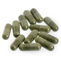 set of identical or nearly identical objects based on the concentration of culturable fungi recovered. Such an item might be an industrial part, a pharmaceutical active ingredient or formulated preparation, or a biomedical device, where items from the same or different lots are compared. This test is generally not useful for indoor environmental investigations.
Field collection method
There is no specific field collection method for this test, per se. Items to be tested should be placed individually in a labeled tube or envelope to which a label containing all relevant sample information has been affixed. The test is semi-destructive, since depending on the size of the specimen, we may need to dissect and subsample it in the laboratory to obtain a suitably sized portion for analysis.
Laboratory code: FC-B
Service options

Results reporting
Results of this test are reported in Colony Forming Units per gram of material (CFU/g), and broken out according to species, species-group or genus observed (the level of identification depends on the taxon in question).
Opportunistic fungal pathogen screen [OFPS]
Published: July 8th, 2009
Revised: March 7th, 2023
Serious infections caused by filamentous or dimorphic fungi are rare in immunologically normal people and normally only caused by one of several pathogenic species. In people with varying degrees of immune dysfunction, however, serious fungal infections are common, and may be caused by a much larger set of fungi many of which are not pathogenic under normal circumstances. These fungi are sometimes called “opportunistic pathogens”, in reference to their proclivity to invade tissues given the opportunity presented by immune compromise. While not typically a problem in the broad community, opportunistic fungal pathogens can be important agents of serious and even fatal disease in health care institutions.
Culture-based methods are the current gold-standard for the detection of opportunistic fungal pathogens, and 1 CFU/m³ has been advised as an action limit (Mayhall 2003, p 706). This test is read rapidly (after 48 hr of incubation). It relies on culture-dependent evaluation of the presence of the most commonly occurring airborne agents of opportunistic fungal disease. Results are reported on the same day that the samples are read.
Taxa included
- Aspergillus species complexes (including A. flavus, A. fumigatus, Aspergillus nidulans, A. niger, A. terreus, and A. ustus)
- Dematiaceous fungi with opportunistic potential (including Curvularia species and relatives, Cladophialophora species, Exophiala species and others)
- Fusarium species complexes (including F. solani, F. dimerum, F verticillioides, and F. oxysporum)
- Paecilomyces species (including P. variotii and Purpureocillium lilacinus species complexes)
- Phaeoacremonium species
- Phialemonium species
- Scedosporium species (including S. apiospermum and S. prolificans species complexes)
- Thermotolerant zygomycetes (including Absidia, Cunninghamella, Mucor, Rhizomucor and Rhizopus species)
- Trichoderma species (including T. citrinoviride/ longibrachiatum complex)
- Yeasts (including Trichosporon, Candida and Cryptococcus species)
Field sample collection
- This test requires the use of an instrument that permits a high sampling flow rate. Instruments including the RCS High Flow, the Andersen N6 and the SAS sampler are suitable, however validation studies have so far only been completed on the Andersen N6. The test requires the collection of a total of 1 cubic metre of air on tandemly collected samples which together are considered as a single sample for the purposes of evaluating the airborne fungal concentration against the target action level of 1 CFU/m³. A proprietary sampling medium is used for this test, which we are pleased to provide to clients.
- Prior to use, the sampling head and exterior of the device should be disinfected with 70% isopropyl alcohol and allowed to dry. This treatment will help to prevent carry-over contamination from previous usages. As well, a clearly-defined sampling plan should be constructed before beginning sampling.
- Verify that the expiration date on the sampling medium is current, and that there is no evidence of damage to the surface of the collection medium (e.g. drying or cracking, colour changes, small glistening, pasty or fuzzy dots that might be microbial colonies, or free water on the medium surface)
- Insert the sampling medium into the air sampler following manufacturer’s recommendations. Wear latex or nitrile gloves during this process to prevent handling contamination.
- Turn the sampler or sampling pump on to initiate air flow. A maximum air volume of 200 L on a single sample should be collected in as many tandem samples are required to sample an overall volume of 1,000nbsp;L of air. All other aspects of this test are similar to IAQ analysis, fungal culture [FC-A]
- After the sampling period has concluded, carefully remove the sampling medium from the device, taking care not to touch the surface of the medium. For Petri plate media, replace the lid and secure it around the edge with tape or Parafilm. Ensure that you use a water-impermeable marker to label the strip, since the strip cannot be identified if the markings become accidentally wetted. Preprinted self-adhesive stickers work well for sample labeling purposes. Be sure to note the order in which samples were collected, and the sample sets that comprise a single collection.
- Place samples in a reclosable clean plastic bag and keep in darkness until receipt at the laboratory. Do not store or ship samples on freezer packs, since this leads to condensation on the agar surface which can lead to the formation of spurious satellite fungal colonies, artifactually inflating the count. Samples need to be protected from abrupt temperature changes during shipment, so it is a good idea to keep them in an insulated box during sampling. The bag containing sample media may be wrapped in bubble wrap or paper and packed into a box or sturdy envelope for shipping. Samples must be received at the laboratory no later than 24 hr post sampling. Please ensure that you use a reliable courier service to meet these requirements. In the past we have experienced numerous problems with Purolator in this regard, and we recommend our clients to use FEDEX for the shipment of time critical samples.
Laboratory code: OFPS
Service options

IAQ analysis, quantitative fungal culture [FC-A]
Published: July 8th, 2009
Revised: March 21st, 2023
Culture-dependent air sampling remains a mainstay of fungal air sampling. Culture-based methods involve the collection of airborne particles by impaction or centrifugation on the surface of a growth medium. The airborne burden of culturable fungi is then extrapolated based on the colony counts obtained after incubation of the sampled media. Culturable air sampling results are usually expressed in Colony Forming Units per cubic metre air (CFU/m³). Although these techniques cannot be used to evaluate human exposure, they are often useful in detecting the presence of indoor fungal contamination. Unlike spore trap sampling, because counts are derived from actively growing colonies, culture-based air sampling affords an ability to identify recovered colonies to a very low level, often to the species level. Culture-based sampling methods do however have several notable limitations, as outlined below.
Methodological limitations
- Sampling intervals must remain brief to minimize osmotic changes in sampling media during collection. In contrast to more integrative sampling techniques possible in spore trap sampling, the use of brief sampling periods increases the influence of transient airborne spore bursts on sample results.
- Standard growth media are selective in their nature, and may not equally support the growth of all fungi present in the air at a given time. Many commonly occurring airborne fungi have specific nutritional requirements that cannot be satisfied by a single general purpose growth medium. Others may be entirely “non-culturable” (e.g. powdery mildew fungi in the genus Erysiphe and others). The growth media typically recommended for culture-based air sampling are intended to optimize recovery of the most relevant subset of the problem indoor fungi.
- A large proportion of the fungal content of air is non-culturable and thus cannot be detected by this sampling method.
- Long incubation times are often necessary to obtain sufficient growth and sporulation to permit accurate identification (e.g. 7-21 d). Attempts to expedite this process by shortening the incubation time or increasing the incubation temperature tend to bias the outcome by selecting for rapidly-growing fungi or thermophilic species, respectively.
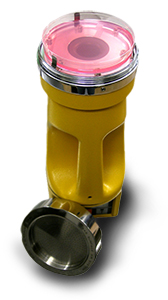
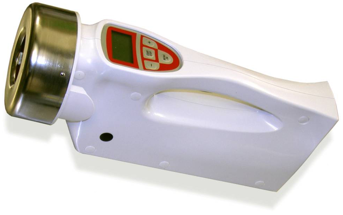 Culture-based sampling is a useful investigative tool to help determine the presence of indoor fungal growth. Culture-dependent sampling is the preferred air sampling technique when low level fungal identifications are required (e.g. to the species-level). This method is also useful where high levels of non-fungal background particulate may be present that would interfere with spore trap testing. Several commercially available sampling instruments can be used for culture-based air sampling, including the RCS Standard and RCS High Flow, Anderson N6 and 2-stage samplers and the Surface Air Sampler (SAS).
Field collection method
-
- Prior to use, the sampling head and exterior of the device should be disinfected with 70% isopropyl alcohol and allowed to dry. This treatment will help to prevent carry-over contamination from previous usages. As well, a clearly-defined sampling plan should be constructed before beginning sampling.
- Verify that the expiration date on the sampling medium is current, and that there is no evidence of damage to the surface of the collection medium (e.g. drying or cracking, colour changes, small glistening, pasty or fuzzy dots that might be microbial colonies, or free water on the medium surface)

RCS YM strip (above), containing a proprietary Rose Bengal Agar
- Insert the sampling medium into the air sampler following manufacturer’s recommendations. Wear latex or nitrile gloves during this process to prevent handling contamination.
- Turn the sampler or sampling pump on to initiate air flow. Flow rates of most units are predefined by the manufacturer of the device. These tend to be in the range of 25-60 L/min. The duration of sampling is usually the only user-controlled variable. During summer months, sample duration should be reduced avoid given the normally high outdoor levels of phylloplane (plant leaf surface) fungi. A final sampling volume of 80-100 L is usually sufficient for most summertime purposes. In the winter, the sampling interval may be doubled unless there is a basis to expect high airborne fungal levels. It is generally not recommended at any time to sample air volumes larger than 200 L on a single sample.
 After the sampling period has concluded, carefully remove the sampling medium from the device, taking care not to touch the surface of the medium. For Petri plate media, replace the lid and secure it around the edge with tape or Parafilm®. In the case of a sampling strip (e.g., RCS), replace the strip in the transport sheath in the same orientation it was removed (e.g. the surface of the growth medium should project into the “dome” inside the sheath — think of it a bit like an ice cube tray). Replacing the strip into the sheath upside down will invalidate the result and the sample cannot be analyzed. Seal the edges of the strip with tape to prevent drying. In all cases, label the sample sheath with the job number, sample number and collection date. Ensure that you use a water-impermeable marker to label the strip, since the strip cannot be identified if the markings become accidentally wetted. Preprinted self-adhesive stickers work well for sample labeling purposes.
- Place samples in a reclosable clean plastic bag and keep in darkness until receipt at the laboratory. Do not store or ship samples on freezer packs, since this leads to condensation on the agar surface which can lead to the formation of spurious satellite fungal colonies, artifactually inflating the count. Samples need to be protected from abrupt temperature changes during shipment, so it is a good idea to keep them in an insulated box during sampling. The bag containing sample media may be wrapped in bubble wrap or paper and packed into a box or sturdy envelope for shipping. Samples must be received at the laboratory no later than 24 hr post sampling. Please ensure that you use a reliable courier service to meet these requirements. In the past we have experienced numerous problems with Purolator in this regard, and we recommend our clients to use FEDEX for the shipment of time critical samples.
Laboratory code: FC-A
Service options

Results reporting
Results of this test are reported quantitatively per taxon identified in Colony Forming Units per cubic metre of air (CFU/m³). Of the microfungi commonly observed in culturable air samples, we generally identify taxa to the species- or species-group level, or to genus-level, wherever is possible within our routine analytical constraints. For example, typically we are able to report most species of Aspergillus to the species-group level (e.g. Aspergillus ustus group, which includes the morphologically very close species A. ustus in the strict sense, A. puniceus, A. panamensis and A. deflectus that cannot be readily differentiated one from the other without specialized procedures). Other taxa that can regularly be reported to the species-group level include Acremonium, Cladosporium, Eurotium, Fusarium, Paecilomyces, Scopulariopsis and Stachybotrys.
By contrast, we routinely identify members of the genus Penicillium to the subgenus-level. This is because identifying species within this group is extremely difficulty in routine practice, and cannot reliably be undertaken by someone who lacks extensive research expertise in this group. Although many environmental mycology laboratories routinely report species-level identifications for this genus, these identifications are dubious at best and cannot be relied upon for any critical purpose. Mostly, they are based on morphological and/ or abbreviated physiological tests given in early monographs, such as Pitt (1979), or less comprehensive treatments including many of the commonly used identification manuals for indoor fungi, including Flannigan et al. (2001), Howard (2003) and Samson et al. (1996) [FULL REFERENCES]. Filamentous fungi that do not sporulate in culture or otherwise produce morphologically distinguishing features are reported as STERILE MYCELIUM. Non-filamentous fungi are reported as yeasts. Other levels of reporting detail are available on an as-need basis for special projects.
WallChek sample [WC]
Published: July 8th, 2009
Revised: March 24th, 2023
The WallChek sampler is a minimally invasive way to test wall cavities for the presence of fungal contamination. The device consists of an Air-O-Cell® sampling cassette outfitted with a 50 cm sampling tube 2.5 cm in diameter attached to the slit face of the impactor. A hole is drilled into the wall assembly to be checked, and the tube is inserted through the hole into the wall cavity. A sampling pump is used to draw air through the cassette at a rate of 15 L/min¹ for 30 sec to 1 min during which time the wall may be gently tapped to release any adherent spores from interior surfaces.
Field sampling method
Click here for operating instructions >
Laboratory code: WC
Service options

Results reporting
Results of WallChek® samples are reported semiquantitatively by fungal group observed, as 1+, 2+, 3+, etc., where the numeral preceding the plus sign indicates the order of magnitude of particles observed in an average 600× or 1000× microscopic field in the central portion of the trace. Background levels are typically very high in WallChek® samples. Nevertheless, they may provide useful information on the condition of the interior of a wall assembly without excessive disruption and minimal risk to investigators and occupants. Quantitative results cannot be provided for this sample type.

